Tag: syringomyelia

  • Brain Under Pressure – A Guide to Understanding Intracranial Hypertension [Updated]

    Brain Under Pressure – A Guide to Understanding Intracranial Hypertension [Updated]

    INTRACRANIAL HYPERTENSION (IH) MEANS HIGH PRESSURE INSIDE THE SKULL.

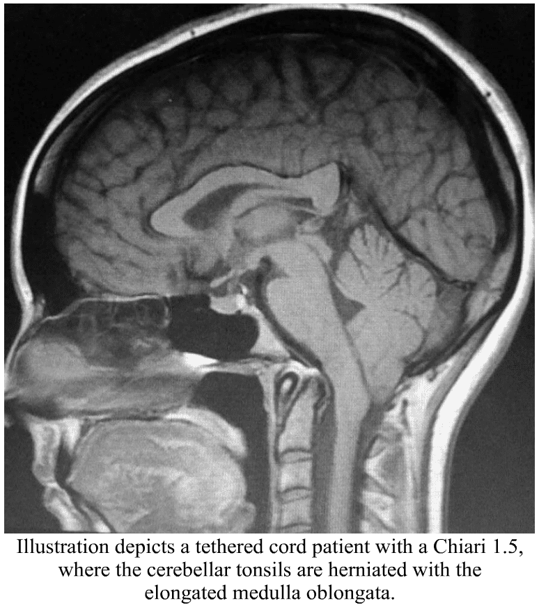
    Intracranial Pressure (ICP) is measured in millimeters of mercury (mmHg). Most scholars agree that on average, “normal pressure” should be between 5-15 mmHg, mild to moderate intracranial hypertension between 20-30 mmHg (which “requires treatment in most circumstances”), and an ICP of > 40 mmHg indicates “severe and possibly life-threatening intracranial hypertension.” [1] When high intracranial pressure is left untreated, it creates a “pushing effect” towards the only natural escape at the base of the skull (the foramen magnum), and the cerebellar tonsils in the pathway are pushed through the foramen magnum. [2]

    Understanding the Monro-Kellie Doctrine (pressure-volume relationship)
    The association between IH/IIH and Chiari Malformation appears to be a malicious intricate pathological circle. The cranium (skull) consists of brain matter, cerebrospinal fluid, and both venous and arterial blood. A hypothesis, referred to as the Monro-Kellie Hypothesis (now better known as the Monro-Kellie Doctrine), states, “The sum of volumes of the brain, CSF, and intracranial blood is constant. An increase in one should cause a decrease in one or both of the remaining two.” Therefore, if an abundance of cerebrospinal fluid (IIH or hydrocephalus), both cranial blood volume and brain matter should be forced to deplete. This depletion is usually directed in the path of least resistance – through the foramen magnum and into the spinal canal. When the brain matter closest to the bottom of the skull (cerebellar tonsils) is pushed through the foramen magnum and into the spinal canal (an Acquired Chiari Malformation), the tonsils act like a cork and blocks the flow of cerebrospinal fluid (regardless of the size of the tonsillar descent), which in turn, continues to raise intracranial pressure.[3]

    More Symptoms of Intracranial Hypertension

    Venous Hypertension
    When an etiological cofactor exists (such as a space-occupying mass), it is considered Secondary Intracranial Hypertension (SIH); when no other cause was identified, it is known as Idiopathic Intracranial Hypertension (IIH) formerly known as Pseudotumor Cerebri. However, recent studies on the connection between Intracranial Hypertension and Venous Hypertension might put an end to the “idiopathic” theory.

    Oxygen-rich blood travels from the heart to the rest of the body through the arterial system, then the oxygen-depleted blood returns to the heart through the venous system. We have a host of small veins in our head and they dump into a series of large veins, called sinuses. Dural Venous Sinus Stenosis occurs when there is a narrowing of one or more of the venous sinuses (most commonly seen in the transverse sinuses or transverse/sigmoid sinus junction), which in turn compromises cerebral venous outflow through the jugular vein (stenosis/compression of the jugular vein can also result in elevated intracranial pressure [4]). Transverse Sinus Stenosis (TSS) is most common in Idiopathic Intracranial Hypertension (IIH). Depending on the study that you are reading, it is proving to be present in 90-100% of IIH patients [5]. While its connection might sound obscure if you look at it from a Monro-Kellie perspective – The blood going into the head, cannot get out at the same speed (because of the narrowed sinus). When this inflow of blood remains constant and the outflow is hindered, the transverse sinus on that side (we have two transverse sinuses, one on each side) enlarges, forcing the CSF and brain matter to reduce to maintain the volume equilibrium. This reciprocation can happen when any of the sinuses or jugular narrow (stenosis). While scholars continue to debate whether TSS is a cause or consequence of IIH, surgeons continue to decompress us without checking our pressures or decompress (the most invasive treatment) in hopes that it will lower our pressures, and patients are left with untreated high pressure still causing a “pushing down effect” and an enlarged foramen magnum for our brains to be pushed down. [2] The sagging brain once again obstructs the flow of cerebrospinal fluid by plugging the foramen magnum, and that in turn raises the intracranial pressure even more. Or, the untreated high pressure blows through the duraplasty and causes a post-operative leak, known as a pseudomeningocele.

    Reducing the Risks of Post-Op IH/IIH Complications
    Brain MRIs often show indicators of Intracranial Hypertension (IH/IIH), therefore, we recommend that all Chiari patients have full brain MRIs and not just cervical MRIs.

    • When the pressure builds inside of the dura mater the pressure pushes the dura and fluid inside of the crevice that holds the pituitary gland (the sella turcica or pituitary fossa). When the amount of fluid is equal to or greater than 50% and the pituitary gland size is 2mm, the condition is known as Empty Sella Syndrome. (Doctors now recognize that < 50% (where the pituitary gland size is 3-7mm) can also cause symptoms and they now refer to that as a partially empty sella.) [8]

    • Slit like or flattened lateral ventricles from the increased pressure, however, when the Foramen of Monro (the aqueduct that connects the lateral ventricle to the third ventricle) is stenosed, the fluid will back-up and the lateral ventricle will not appear flattened. [7]

    • Enlarged/swollen optical nerves (papilledema). [8]

    • Low lying or herniated tonsils (often diagnosed as a Chiari Malformation). [2]

    What We Recommend BEFORE DECOMPRESSION is considered:
    If you have symptoms of IH/IIH accompanied by any of the MRI indicators mentioned above, it is both reasonable and prudent to ask your neurosurgeon to investigate further BEFORE DECOMPRESSION.

    • See a neuro-ophthalmologist to check for signs of papilledema, including Optical Coherence Tomography and Ultrasonographic B-scanning. [8]
    • Magnetic Resonance Venography (MRV, preferably with the ATECO technique) to check for venous stenosis of any of the cranial sinuses and/or jugular vein. Stenosis is not exclusive to the transverse sinus and it can happen in multiple sinuses simultaneously.
    • If overweight, consider trying to lose weight. Studies show that a weight loss of 5-10% of one’s overall body weight, when accompanied by a low-salt diet, can offer some to IH/IIH symptoms.[9]
    • Consider trying Diamox (Acetazolamide) and/or Topamax (Topiramate) to see if that improves the pressure headaches.
    • Request a lumbar puncture (spinal tap) to test your opening pressures. We recommend that it’s guided with fluoroscopy with a small gauge needle (and not the standard 22 gauge) that they allow to drip (as opposed to syringe pull) and ensure that someone is available to perform an epidural blood patch if necessary. Time should be allotted afterward to lay flat for several hours immediately following the procedure and for several days once returning home. The potential for CSF leaks is high for the EDS/Chiari patient. A doctor that marginalizes the risks ahead of time, will generally marginalize your symptoms when you are actively leaking.
    • ICP Bolt Monitoring can record the differences experienced in pressure over time, and how different positions affect ICP.

    Note: When the intracranial pressure gets high enough, it can cause a cranial leak. This is especially true for the Ehlers-Danlos patient where the dura mater is thin and fragile. When a cranial leak decreases the intracranial pressure, the papilledema, empty sella, stenosis, and high-pressure headaches can sometimes start to revert to normal or near-normal, and the leak will affect any attempts to check intracranial pressure (reducing the pressure from what it was before the leak occurred), however, the tonsillar herniation will usually remain if the pressure gets too low. [10]

    TREATMENT OPTIONS:
    If Venous Stenosis exists, stenting should be considered as leaving the sinus/jugular stenosed can post other health risks, and stenting is proving to have much better success with fewer complications requiring revisions. When medication fails to decrease ICP, and a stent is not an option, a Ventriculoperitoneal Shunt (VP Shunt) or Ventriculoatrial Shunt (VA Shunt) can be surgically placed to drain cerebrospinal fluid straight from the ventricle. Shunts are known for failing and often need a multitude of revisions, but even with all the revisions, it is less invasive than a decompression. Shunts under the foramen magnum should never be used as a means of controlling ICP.

    For the IH/IIH patient, herniated tonsils should be assumed an Acquired Chiari Malformation (even if a small posterior fossa is evident), and by correcting the high pressure before decompression, the decompression will be less likely to fail.

    Helpful Tips:
    If you have IH/IIH, it is best to avoid caffeine, avoid progestin based birth control, and all EDS patients should try to avoid the use of fluoroquinolones such as ciprofloxacin (Cipro), levofloxacin (Levaquin/Quixin), gatifloxacin (Tequin), moxifloxacin (Avelox), ofloxacin (Ocuflox/Floxin/Floxacin), norfloxacin (Noroxin), due to the increased risk of aneurysm.

    [wpedon id=”4396″ align=”center”]


    References: 

    1 Rangel-Castillo, Leonardo, et al. “Management of Intracranial Hypertension.” Rangel-Castilla, Leonardo et al. “Management of intracranial hypertension.” Neurologic clinics vol. 26,2 (2008): 521-41, x. doi:10.1016/j.ncl. Feb. 2008, <https://www.ncbi.nlm.nih.gov/pmc/articles/PMC2452989/>.

    2 Aiken, A.H., et al. “Incidence of Cerebellar Tonsillar Ectopia in Idiopathic Intracranial Hypertension: A Mimic of the Chiari I Malformation.” American Journal of Neuroradiology; Nov. 2012, <http://www.ajnr.org/content/33/10/1901>.

    3 Mokri, B. “The Monro-Kellie Hypothesis: Applications in CSF Volume Depletion.” Neurology., U.S. National Library of Medicine, 26 June 2001, <https://www.ncbi.nlm.nih.gov/pubmed/11425944>.

    4 Zhou, D., et al. “Intracranial hypertension induced by internal jugular vein stenosis can be resolved by stenting.” European Journal of Neurology, November 2017 <https://onlinelibrary.wiley.com/doi/abs/10.1111/ene.13512>.

    5 Henderson, Fraser C., et al. “Neurological and Spinal Manifestations of the Ehlers–Danlos Syndromes.” American Journal of Medical Genetics Part C: Seminars in Medical Genetics, 21 Feb. 2017, <www.onlinelibrary.wiley.com/doi/10.1002/ajmg.c.31549/full>.

    6 Pietrangelo, Ann. “Empty Sella Syndrome.” Healthline, Oct. 2017, <https://www.healthline.com/health/empty-sella-syndrome>.

    7 Hingwala, Divyata R., et al. “Imaging signs in idiopathic intracranial hypertension: Are these signs seen in secondary intracranial hypertension too?.” Annals of Indian Academy of Neurology vol. 16,2: 229-33. doi:10.4103/0972-2327.112476, June 2013, <https://www.ncbi.nlm.nih.gov/pmc/articles/PMC3724081/>.

    8 Mollan, Susan P., et al. “A practical approach to, diagnosis, assessment and management of idiopathic intracranial hypertension.” Practical neurology vol. 14,6: 380-90. doi:10.1136/practneurol-2014-000821. May 2014, <https://www.ncbi.nlm.nih.gov/pmc/articles/PMC4251443/>.

    9 Thurtell, Matthew J., and Michael Wall. “Idiopathic Intracranial Hypertension (Pseudotumor Cerebri): Recognition, Treatment, and Ongoing Management.” Current Treatment Options in Neurology, U.S. National Library of Medicine, Feb. 2013, <https://www.ncbi.nlm.nih.gov/pmc/articles/PMC3554852/>.

    10 Pérez, Mario A., et al. “Primary spontaneous cerebrospinal fluid leaks and idiopathic intracranial hypertension.” Journal of neuro-ophthalmology : the official journal of the North American Neuro-Ophthalmology Society vol. 33,4: 330-7. doi:10.1097/WNO.0b013e318299c292, Dec. 2014, <https://www.ncbi.nlm.nih.gov/pmc/articles/PMC4040082/>.

  • Spine Pulled Tight – A Guide to Understanding Tethered Cord Syndrome

    Spine Pulled Tight – A Guide to Understanding Tethered Cord Syndrome

    TETHERED CORD SYNDROME (TCS) INVOLVES A STRETCHING OF THE SPINAL CORD, AND OFTEN YOUR MEDULLA OBLONGATA
    AS WELL, WHICH LEADS TO A HOST OF NEUROLOGICAL PROBLEMS.


    Before we talk about Tethered Cord Syndrome, let’s first talk about the anatomy associated with the spinal column (in layman’s terms).

    •  The role of the vertebral column is to hold the spine strong (so it can be upright) and protect the spinal cord from injury. In a normal vertebral column, there are thirty-three vertebrae on each side (seven cervical vertebrae, twelve thoracics, five lumbar, five fused vertebrae in the sacrum and another four fused vertebrae in the coccyx).

    •  Each vertebra in the upper twenty-four vertebrae is separated by intravertebral discs largely composed of the fibrous protein, collagen. The main role of these discs is to allow the vertebral column to move and flex.

    •  The role of the spinal canal is to hold cerebrospinal fluid around the spinal cord, which not only cushions the cord against injury, but it also lubricates the cord, cleanses the cord, and brings essential nutrients that the spinal cord needs. The spinal canal is made up of several layers that form the meninges. These layers are also composed of high concentrations of collagen. The outermost layer of the meninges is the dura mater. The dura mater should be dense and strong, so cerebrospinal fluid cannot leak from it.

    •  The spinal cord relays messages from the brain to the nerves that allow the body to function. When part of the spinal cord is compromised, it can inhibit signals from getting to the nerves from that point downward. The brainstem (midbrain, pons, and medulla oblongata) is attached to the spinal cord at the top (the medulla and spinal cord meet at what is called the cervicomedullary junction) and the spinal cord continues down to the mid/lower back. From there it becomes a delicate elastic band of fibrous collagen-based tissue called the filum terminale that extends from the conus medullaris (the lowest point of the spinal cord before it becomes the filum terminale) to the dural sac at the S2 level.1

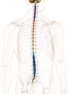
    Chiari Malformation has many conditions that can be associated with it (comorbid conditions) and sometimes those comorbid conditions can be at the root cause (etiological cofactor) or one of the causes along the way (pathological cofactor) to the tonsils being as low as they are (making the Chiari “secondary” to one or more “other” conditions). Tethered Cord Syndrome (TCS) is one of those pathological conditions.2 Like Chiari, it is a neurological disorder; however, it is one of the spinal cord.3

    Tethered Cord happens when the sticky fibrous tissue of the filum adheres to fatty/scar tissue or the dura lining of the spinal canal.1 While this tethering can happen anywhere in the spinal canal, it is most common at the lumbosacral level.4 When the tethered filum pulls the spinal cord tightly enough that it causes neurological problems, it becomes known as Tethered Cord Syndrome (TCS). Tethered Cord is most common in patients with Spina Bifida (myelomeningocele, meningocele), Spina Bifida Occulta (lipomeningomyelocele, lipomyelocele) and patients with Ehlers-Danlos Syndromes (EDS), a Hereditary Disorder of Connective Tissue (HDCT) where one or more of the types of collagen (the most abundant protein in the human body) is mutated at a cellular level. Tethered cord can be congenital or acquired. It can be obvious in childhood or symptoms may not present themselves until adulthood. Some children may develop minor signs that are overlooked by untrained medical professionals and can progress slowly or rapidly over time.

    More Symptoms of Tethered Cord Syndrome

    A Tethered Cord Syndrome diagnosis can be somewhat of a challenge. The signs and symptoms of the condition are not always present and when they are, they are often not recognized, so it is important to know all indicators. People with Tethered Cord (TC) can have sacral dimples, discoloration, and hairy patches on their lower back that can lead a doctor to investigate further, however, some have no external signs at all. Some have kyphosis (rounded back) and scoliosis (curved spine). Sometimes radiological criteria are not met or are ambiguous, yet an Occult Tethered Cord (characterized by the presence of symptoms with normal conus position and inconclusive findings of the filum) can still exist.5 Symptoms can be elusive as well and can happen all at once or gradually over the course of many years. Many symptoms worsen due to activity; climbing stairs has been reported as causing pain that varies from uncomfortable to excruciating.

    One of the reasons that Tethered Cord is often overlooked is that many neurosurgeons are not aware of the connection it can have with a Chiari Malformation and the medical tests used for determining if a tethered cord problem exists are not always accurate or accurately read.


    Magnetic Resonance Imaging (MRI)

    • A lumbar MRI is usually the first step. This gives a visualization of the spinal cord in relation to the surrounding vertebrae. The actual tethering is not always obvious on MRI, sometimes the only proof of the tethering is the pulling it creates on the spinal cord. Doctors will look for the position of the conus medullaris when looking for signs that the spinal cord is being pulled. The consensus amongst most surgeons is that a normal conus should be located from the T12 to the lower L2. There is much debate on the importance of establishing evidence of a low-lying conus.5 When conus reaches the lower L2 or below, doctors should be investigating why it’s low and consider if the cord might be tethered. When looking for the location of the conus, your position can make all the difference. MRIs are generally performed supine (lying down) and the cord is not pulled as tightly as it is when upright. For this reason, upright MRIs are becoming the method of imaging preferred by most neurosurgeons looking to confirm or deny if tethering exists in a patient showing symptoms. Other signs of tethering that might be visible in a lumbar MRI include an enlarged foramen magnum, thick or fatty filum, presence of fatty tissue inside the canal, or the filum might be pulling to one side of the canal.5

    • A prone MRI of the lumbar region can be an invaluable tool for those where other MRIs indicate that the filum might be pulling to one side (usually the back side) of the canal. With prone MRIs, imaging is done while the patient is lying face down (as opposed to facing up, like most supine MRIs). If the anteroposterior conus movement of >10% of the canal width was evident from the supine to the prone, then the likelihood of it pulling to one side do to tethering is less likely and more conservative management might be better appropriate than a surgical release.6

    • A cervical MRI can also show signs that a tethered cord might exist. The cervical spinal cord can sometimes appear narrow from it being pulled tight. The medulla oblongata can become elongated. This happens because the brainstem is attached to the top of the spinal cord and that cord is being pulled tight, essentially pulling everything down and tight. This elongation of the medulla from the tethering can cause secondary symptoms by itself, known as Dysautonomia.

    · Low/herniated cerebellar tonsils consistent with what is seen in a Chiari Malformation. When the brainstem is herniated (where part/most of the medulla is below the foramen magnum) along with the cerebellar tonsils, it is considered a Chiari 1.5 (which should be a good indicator that you might be dealing with an Acquired Chiari Malformation, where the herniated tonsils are secondary to another condition). One study quoted that out of 2,987 patients with a tonsillar herniation of 5mm or greater, 14% met the diagnostic criteria (based on “generally accepted clinical and radiographic criteria”) and 63% of the 289 patients with tonsillar herniations of < 5mm.5

    · A syrinx is common with Tethered Cord, as it causes a blockage of fluid at the foramen magnum. A syrinx can develop anywhere in the spine, usually in the lower cord, but with Tethered Cord Syndrome it can develop in the lower medulla (Syringobulbia) as well because of the low brainstem is at the point of the blockage of fluid from the Chiari Malformation.

    Even with an upright MRI and every symptom listed, patients are often told they do not have Tethered Cord. This is simply due to a lack of education on the subject and medical bias between doctors. It is important to make sure that you have the images viewed by a neurosurgeon that is familiar not only with Tethered Cord but Chiari and Comorbids as well. (Nearly half of the large study quoted above were referred following a failed Chiari Decompression.5) The combination of the images and the patient’s symptoms should tell the neurosurgeon if surgical intervention is required. Patients often require several consultations before they can find a knowledgeable enough physician.


    What We Recommend BEFORE DECOMPRESSION is considered:
    If you have symptoms of TCS, especially if accompanied by any of the MRI indicators mentioned above, it is both reasonable and prudent to ask your neurosurgeon to investigate further before decompression is considered. A Tethered Cord Release Surgery prior to decompression may relieve the tension that is pulling the brainstem and cerebral tonsils downwards reducing the risk of a failed decompression. There is a chance with small tonsillar herniations that the Tethered Cord Release might allow the cerebellar tonsils to rise enough to the point that cerebrospinal fluid flow is reestablished to where decompression is no longer needed. However, failure to release a tethered cord prior to decompression surgery increases the likelihood of a failed decompression. (In fact, in the study quoted above, out of the 3,276 patients with herniated tonsils, 46% of them were referred for evaluation after a failed decompression surgery.5) An MRI of all three levels of the spine should be done to rule out other possible causes for leg/back symptoms along with urodynamic testing, an Electromyogram (EMG) Test and Nerve Conduction Study (NCS) of the lower limbs is also suggested.


    TREATMENT OPTIONS:
    For some, physical therapy can help with symptoms for a while. However, ultimately it will likely need to be surgically treated with a Tethered Cord Release.

    Tethered Cord Release (TCR) Surgery involves the untethering of the spinal cord. An incision is made in the lumbar area, the filum terminale is separated and the factors that are tethering the spinal cord to the vertebrae are severed. Surgical treatment is not without risk and does not guarantee relief of symptoms. However, in a large study, up to 83% of adult patients report relief, 16% unchanged, and 1% report feeling worse.5 In children, the numbers are even better with 93% obtaining improved symptoms and 7% unchanged.5 Most patients describe the surgery as extremely painful for the first two weeks and “better than they ever remember feeling” (often because they have been tethered for much of their lives) after two weeks. The most common complication involves retethering (often from the scar tissue from the release) and multiple surgeries may be required over a lifetime. Finding a neurosurgeon experienced with TCRs and the surgical treatment of Ehlers-Danlos patients can sometimes help reduce the risks associated with scar tissue formation, but scar tissue can happen with even the best of neurosurgeons.

    For the TCS patient, herniated tonsils really should be assumed an Acquired Chiari Malformation (even if a small posterior fossa is evident), and by correcting the tethered cord before decompression the decompression will be less likely to fail.


    Special Note: There are other conditions that can present with similar symptoms. Diastasis Recti is a type of abdominal hernia common to pregnant women, those with obesity, and EDS patients. This separation in the abdominal muscles is known to sometimes cause lower back pain and many of the same pelvic floor problems seen with Tethered Cord Syndrome (TCS). Unlike TCS however, it does not usually require surgical treatment. If you suspect Diastasis Recti, we recommend that you talk to your Primary Care Physician about referring you to physical therapy to bring your abdominal muscles back together before considering Tethered Cord Release (TCR).7


    [wpedon id=”4396″ align=”center”]


    References: 

    1 Henderson, Fraser C., et al. “Neurological and Spinal Manifestations of the Ehlers–Danlos Syndromes.” American Journal of Medical Genetics Part C: Seminars in Medical Genetics, 21 Feb. 2017, <www.onlinelibrary.wiley.com/doi/10.1002/ajmg.c.31549/full>.

    2 “Section of the Filum Terminale Surgery for Tethered Spinal Cord Syndrome in Patients with Chiari Malformation and Syringomyelia.” North Shore-Long Island Jewish Health System The Chiari Institute, Chiari Connection International, 02 Oct. 2006, <http://www.chiariconnectioninternational.com/docs/TCS_SFT_Explained.pdf>.

    3 Quake. “Overview: Chiari Comorbidities & Etiological/Pathological Cofactors [Revised].” Chiari Bridges, 16 Nov. 2019, <http://chiaribridges.org/overview-chiari-comorbidities-etiological-pathological-cofactors/>.

    4 Arslanoglu, A., et al. “Multidisciplinary Combined Approach for Tethered Spinal Cord Syndrome: Radiology, Surgery and Physical Therapy.” Balkan Military Medical Review, 2006, <https://pdfs.semanticscholar.org/8c30/18bf5bfbd6f3e7e5e9c3559bbbfdcac82e04.pdf>.

    5 Milhorat, Thomas H., et al. “Association of Chiari malformation type I and tethered cord syndrome: preliminary results of sectioning filum terminale.” Surgical Neurology, Elsevier, July 2009. <https://www.sciencedirect.com/science/article/abs/pii/S0090301909002572>.

    6 Aoun, Salah G., et al. “The Use of Prone Magnetic Resonance Imaging to Rule Out Tethered Cord in Patients With Structural Spine Anomalies: A Diagnostic Technical Note for Surgical Decision-making.” Cureus vol. 11,3 e4221. 11 Mar. 2019, doi:10.7759/cureus.4221. <https://www.ncbi.nlm.nih.gov/pmc/articles/PMC6510567/>.

    7 “A Complete Guide to Diastasis Recti: Truths on Abdominal Muscle Separation.” Braceability, 05 June, 2018. <https://www.braceability.com/blogs/info/diastasis-recti>.

  • The Sarah Taylor Story – A Chiari Warrior’s Journey

    The Sarah Taylor Story – A Chiari Warrior’s Journey

    When I first started getting hit with symptoms, I was a divorced, single mother of three amazing kids; responsible not only to provide for them but to see them through life, unscathed by life’s situations, and showing them that there was nothing that if they worked hard at something, nothing could hold them back. I had just started to expand in my career as a self-taught auto technician. I was a woman making a place for herself in an industry traditionally dominated by males. July 3, 2015, was the day that my life forever changed. I was brought to the hospital with stroke-like symptoms. I was having visual problems. I couldn’t walk or talk. I had no idea who I was or where I was. The whole right side of my body basically stopped working and the right side of my face was droopy. I was brought to the ER and before the doctor would even try to figure out what was wrong with me, he ordered a series of drug tests. I passed every test, so he finally admitted to me. Once on the neurology floor, more testing was done. They performed an MRI, MRA, EKG and told us that all results were normal. I later discovered that was not the case.

    One doctor refused to believe that I was not on drugs. She noted in my file that while she has no evidence to support it, she believed that I am on a drug that they hadn’t screened for, based solely on “my age, single mom status, and prior good health.” She also noted that they found a Chiari, but that based on my symptoms, she believed it was irrelevant (an incidental finding). I would love to see her now and show her just how very wrong she was. I firmly believe that what she put in my medical chart is why I have had such a difficult time getting the care that I need and deserve.

    A few days later, I followed up with my PCP. She went over my MRI results with me and pointed out that they found a Chiari Malformation with a 19mm herniation of my cerebellar tonsils. She told me of changes in my white matter that the radiologist said needed to be “further evaluated” and referred me to my first neurologist, who I met within August. He ordered a visual evoked potential and an EEG. Both come back normal, so he diagnosed me with migraines, even after hearing my symptoms, which frustrates me even more as I know that it is not migraines causing these issues.

    At this point, I switched my neurology care to another hospital. They went over my history with me and ordered a lumbar puncture to rule out Multiple Sclerosis, which showed banding in my spinal fluid. On September 14, 2015, I was officially diagnosed with Multiple Sclerosis and opted to begin treatment and was to start on Plegridy. As I started the full doses I started breaking out into hives. The docs didn’t seem to think I should worry, so I called the drug manufacturer and they said it should be considered an allergic reaction to the Plegridy, and to discontinue using it and advise my doctor.

    After this experience, I switched care back to the first hospital for neurology to get a second opinion. The new neurologist ordered a new brain MRI and one of my cervical spine. There were no changes to my brain MRI, but my cervical imaging showed a syrinx. They weren’t sure if the syrinx was of any significance. So, she referred me to the only MS specialist in North Dakota whom I would meet with, in May. The MS specialist took a complete history on me and ran a bunch of blood work to rule out other illnesses. When those illnesses were all ruled out, she diagnosed me with Radiologic Isolated Syndrome (which means that they saw similar characteristics to MS in my imaging, without MS symptoms). While in her care I continued to get worse, with symptoms progressed to include pins and needles feeling in my hands and feet, occipital headaches that drop me to the ground, cognitive decline, fatigue, weakness, some random numbness, and muscle spasms. She ordered a new MRI and once again no changes were indicated. She began to question if my Chiari was behind my growing number of symptoms. She tried to refer me to Mayo, but my insurance declined her referral.

    Eventually, I started having issues walking and my gait was becoming increasingly unsteady, so I return to a local neurology clinic. They did an MRI on my brain, cervical and thoracic spine. They found a syrinx in my thoracic spine and once again they doubted the significance, along with a slight scoliosis convex. When asked what a syrinx was, they told me that it was “an old MS lesion.” I later learned that a syrinx is a cyst inside of the spinal cord caused by a blockage of cerebrospinal fluid and it damages the spinal cord from the inside out – often associated with Chiari Malformation.

    During this care for MS, I kept having what they thought were MS relapses, roughly every three to four months. Each time they ordered new MRI images and treated me with high doses of IV steroids for five days in a row. Never once did this imaging ever show an actual MS-relapse or MS activity. I continually had issues with every medication that they put me on to help “try to delay the progression of the MS” (the MS that I never had). In November 2017 I started Ocrevus, which was just FDA approved that year. Around this time, I started having strange symptoms and thought them just to be side effects of the medication, not realizing that something else might be causing it all. I met with my neurologist before my second full dose and I told her everything that I was experiencing. We opted to take me off the Ocrevus and they repeated the MRI yet again. Again, the MRIs show absolutely nothing new for activity and she admits that she doesn’t know what to do for me. I am three years in at that point and never once have they seen any MS activity.

    I made an appointment with yet another neurologist. I met with him on March 2019 and he immediately pulled my MS diagnosis. He instead decides that I have migraines and anxiety. He believes that anxiety is why I am completely numb all over my body. He disregards the Chiari and the syrinxes when asked about them stating that they do not cause any symptoms that aren’t of any significance. I left this appointment more frustrated than I was before and began losing hope that I was ever going to be able to figure out what was going on with me. How am I ever going to get the proper treatment when I am consistently blown off whenever I ask about a condition that was noted from day one?

    I began working more closely with my PCP. I went over the last three years of my medical journey with her and told her that I felt that we really needed to dig into this Chiari Malformation that has been called out in my imaging since July of 2015, especially since I had many symptoms that may be from it. I told her about a neurosurgeon that I had been told about in Sioux Falls, SD, who specializes in Chiari. We also talk about a connective tissue disorder known as Ehlers-Danlos Syndrome and start comparing my symptoms (of which I had several). My PCP sends in referrals to the neurosurgeon, a genetic counselor, and a rheumatologist. (Because with Chiari you will more than likely have several comorbidities.)

    In June we traveled down to the specialist. He went over my MRI images and stated that my herniation was 19mm (which was almost quadruple the amount that they get concerned about). That coupled with my symptoms led to discussing the need for me to have decompression surgery. Finally, after four years we know the true culprit of what was wrong with me, my brain is literally falling out of my skull. We leave with a bunch of literature for the surgery and I call his office back Friday and tell them my decision to go forward with the surgery and we started planning for me to have surgery in early September. Just as we thought everything was on the right course, my insurance drops a bombshell on me. I received a call from the specialist office, and they tell me that my insurance has declined my surgery stating that I can have it done locally by the same incompetent neurosurgeon that I met who couldn’t even measure my Chiari correctly. I have appealed this decision twice and both times I was denied. I am now pushing for a State Fair Hearing.

    The last four years have been one hell of a ride when it comes to my health. My health problems have made it far more difficult to continue working on cars. As my symptoms wage war on my body, I am now forced to work on light duty and have been for the last two and a half years. I know that my days of working in a shop are coming to end as I just can’t handle the physical requirements of the job anymore. My quality of life in the last year alone has declined sharply. I used to be the energetic mom who could coach a sports team after working all day in the shop and still have the energy to keep up with the housework, now that is not the case. I manage to push on and get them to their activities, but I’m exhausted to the core. When this all began back in 2015 my kids were 8, 7, and 5. They are now 12, 11 and 9. At times I feel like I am a horrible mother because I miss the mom that I used to be. I miss the days when my kids weren’t worried about my health and when we could make plans with other families and keep them. I have lost so much of who I am thanks to the ignorance of some members of the medical community. I am losing faith in the medical profession in general. Male doctors have been the worst as I go through this journey, as women seem to have to first prove that it’s not psychosomatic before we’re worthy of being helped, even with imaging shows something to the contrary. When I present them with proven facts about Chiari Malformation, it still gets dismissed and it is extremely frustrating. The longer I go without receiving proper treatment, the more likely it becomes that some of this damage will become permanent and to me, that is not acceptable. I am fighting for my life and I will not back down until I receive the proper care, I can’t!

  • Overview: Chiari Treatment Options & Potential Pitfalls

    Overview: Chiari Treatment Options & Potential Pitfalls

    Once diagnosed, you will usually be referred to a specialist (not a Chiari Specialist, but an everyday, run-of-the-mill neurologist or neurosurgeon). They tend to come in one of two types: Either they are very passive and just want to wait and see how bad it gets, or they are very pro-surgery and while they will still usually give you a 50% chance of helping your symptoms, they will tell you how decompression surgery really is your best option. Both are problematic.

    FOR THE ASYMPTOMATIC CHIARIAN:
    Without a doubt, the passive approach is by far the best plan of action for the asymptomatic Chiari patient. Approximately 30% of those with a Chiari 1 malformation can go their entire lives without having symptoms (in fact, many remain undiagnosed and don’t even know they have it because they have no symptoms).[1] The Chiari is often discovered while looking for something else entirely and is therefore considered an “Incidental Finding.” While there is a chance that the Chiarian will become symptomatic one day, the risks of surgical complications exceed the potential benefit on something that has not and might not ever affect their life.

    FOR THE SYMPTOMATIC CHIARIAN:
    If the Chiarian is symptomatic, however, “Decompression Surgery is the only treatment available [at this time] to correct functional disturbances or halt the progression of damage to the central nervous system. Most individuals who have surgery see a reduction in their symptoms and/or prolonged periods of relative stability. More than one surgery may be needed to treat the condition.”[2] Despite the reasons for haste however, we do recommend slowing down and making sure that adequate testing is done to ensure that the Chiari is a “Congenital Chiari” formed only by a small posterior fossa, with no other etiological/pathological co-factors that could make it an “Acquired Chiari Malformation.” If not identified and addressed, these etiological/pathological co-factors can cause complications and even lead to a failed decompression surgery. (Note: most are told at diagnosis that it is a “congenital defect.” However, that is usually a presumption on their part, stemming from a lack of knowledge of Chiari and its comorbidities, and them giving too much credence to the paragraph or two on Chiari malformations in their medical school textbooks. That is not the reality that many are dealing with; so, testing is imperative!)

    TESTING:
    If you have been diagnosed with a Chiari malformation or a tonsillar ectopia (regardless of the size of tonsillar descent), you should have the following tests/images done.

    1. A sleep study to check for Sleep Apnea.

    • Central Sleep Apnea (CSA) is more common with Chiari, especially when there has been damage to the brainstem or Vagus Nerve.

    • Obstructive Sleep Apnea (OSA) is commonly linked with obesity in the general population, it is also very common amongst those with Connective Tissue Disorders (such as Ehler’s-Danlos Syndrome).

    • Both CSA and OSA can be present in the Chiarian with a Connective Tissue Disorder. When both are causing apnea, it becomes known as “Complex Sleep Apnea.”

    • Sleep Apnea, regardless of the type, is a common “killer” amongst Chiarians.


    2. A brain and full spine MRI (upright recommended) with and without contrast.

    What they should be looking for in these MRIs:

    a) Do you have a syrinx (Syringomyelia or Syringobulbia)?

    • If you have either, that is proof that the blockage of CSF is significant enough to cause these potentially serious complications.

    b) Is any part of your brainstem below the foramen magnum?

    • If it is, you have a variant of Type 1, known as a Chiari 1.5[3] or Type 2, both indicate that there is something else going on causing the brainstem to herniate along with the cerebellar tonsils. 

    c) Do you have a cyst/tumor causing increased intracranial pressure that is pushing the cerebellar tonsils down?

    • If you have either, and they are operable, there is a chance that surgically removing it could decrease the intracranial pressure and allow the cerebellar tonsils (and brainstem if it is below the foramen magnum at all) to go to proper position. Furthermore, if the cyst/tumor is not addressed before, during, or soon after decompression surgery, the chances of your tonsils herniating again after decompression are high.[4]

    d) Do you have Hydrocephalus causing increased intracranial pressure that is pushing the cerebellar tonsils down?

    • If you do, there is a chance that the placement of a Ventriculoperitoneal Shunt could decrease the intracranial pressure enough to allow the cerebellar tonsils (and brainstem if it is below the foramen magnum at all) to go to their proper position. Furthermore, if the Hydrocephalus is not addressed before, during, or soon after decompression surgery, the chances of your tonsils herniating again after decompression are high.[5]

    e) Do you have signs/symptoms of Idiopathic Intracranial Hypertension (often shows as excessive fluid above the lateral ventricles, with small, “slit-like” lateral ventricles; usually accompanied by Papilledema and/or Empty Sella Syndrome), which can cause enough cranial pressure that it can push your cerebellar tonsils down?

    • If you do, there is a chance that a prescription for Diamox or Topamax, or the surgical placement of a Ventriculoperitoneal Shunt, could reduce the pressure enough to allow the cerebellar tonsils (and brainstem if it is below the foramen magnum at all) to go into proper position. Studies exist both in favor of shunting first and doing the decompression first; there are documented cases where unresolved IH has led to failed decompression surgeries, while other cases attribute a Chiari decompression as being that which resolved IH symptoms.[6/7]

    f) Do you have signs/symptoms of Tethered Cord Syndrome that could be pulling on the spine from below?

    • If you do, there is a chance that a less invasive surgery known as a Tethered Cord Release, could stop the downward pulling of the spinal cord and allow the cerebellar tonsils (and brainstem if it is below the foramen magnum at all) to go into proper position. Furthermore, if the Tethered Cord is not addressed before, during, or soon after decompression surgery, the chances of your tonsils herniating again after decompression are high. (Note: not all Tethered Cords are easily visible by MRI, when they are not visible, it is called Occult Tethered Cord (OTC) and surgery on a OTC remains controversial.)[8]

    g) Do you have signs/symptoms of Intracranial Hypotension (CSF Leak) that could be pulling or creating a suctioning effect from below?

    • If you do, there is a chance that a less invasive blood patch or a Dural Tear Repair Surgery, could stop the downward pulling/suctioning effect on the spinal cord and allow the cerebellar tonsils (and brainstem if it is below the foramen magnum at all) to go into proper position. Furthermore, if the leak(s) are not addressed before, during, or soon after decompression surgery, the chances of your tonsils herniating again after decompression are high. (Note: not all leaks are easily visible by MRI.)[9]

    h) Do you have signs of Spina Bifida (Myelomeningocele, Meningocele, or Spina Bifida Occulta)?

    • While it is important to know if any of these exist, a Myelomeningocele would put you at an increased risk of having a Chiari Type 2. While all Chiari Malformations with a Myelomeningocele do not meet the standards for Type 2, 90% of those with a Myelomeningocele have a Chiari Type 2. Most neurosurgeons will not perform a decompression on someone with a Myelomeningocele because of the risk of further complications.[10]

    i) Do you have signs/symptoms of disc degeneration problems and/or stenosis, especially in the cervical spine where it can damage the Vagus Nerve?

    • If you do, these could be another sign of a connective tissues disorder. If you have a syrinx above a herniated/bulging disc, there is a good chance that the disc is attributing to it. Herniated/bulging discs in the cervical spine can be quite troublesome. They can usually be addressed by a second procedure called an Anterior Cervical Discectomy and Fusion (ACDF), but if there are multiple cervical problems and Craniocervical Instability, some surgeons will opt to do a fusion at the time of decompression.[11]

    j) Do you have signs and symptoms of a cervicomedullary syndrome[12], which are often the result of Craniocervical Instability (CCI), Atlantoaxial Instability (AAI), or both. (CCI and AAI can be confirmed with: An upright c-spine MRI with flexion, extension, and neutral views or a 3D cervical CT with rotational views.)

    • CCI/AAI can produce many of the same symptoms as Chiari 1 malformation, including occipital or craniocervical junction headaches made worse by Valsalva maneuvers. It has been theorized that CCI/AAI may also be capable of causing or worsening a herniation of the cerebellar tonsils, and it has been demonstrated that CCI can cause ventral brainstem compression and deformative stress injury to the brainstem.[13] Failure to diagnose and treat CCI before or along with decompression has also been linked to decompression failure. CCI and AAI, while rare in the general population, are relatively common in patients with HDCT’s (Heritable Disorders of Connective Tissue), such as Ehlers-Danlos Syndrome. CCI, when identified to be the root cause of significant symptoms, can be treated conservatively, and in the short term, with the use of a hard-cervical collar, physical therapy and, possibly, traction. More permanent treatment in the form of a fusion and stabilization surgery (fusing the occiput to C1, C2, and sometimes additional vertebrae).[12/13]

    3. A preliminary check for connective tissue disorder (especially Ehler’s-Danlos Syndrome). If preliminary results indicate the presence of a connective tissue disorder, genetic testing should be done to rule out more serious types.


    IF NONE OF THESE PROBLEMS APPEAR TO EXIST: 

    Some doctors and patients prefer to wait and just treat the symptoms medically. That is your choice to make and you should never let anyone try to bully you or manipulate you into having surgery or not; although it is important to know that statistics show that those who have had a shorter duration of onset of symptoms and surgery tend to have a better surgical outcome.[14]

    Statistics show that 80% of decompressed patients report some relief from some of their symptoms; headaches are the most commonly reported symptom to gain relief (81%).

    Most surgeons will give a 50% chance of relieving each symptom individually, with the exception of pain associated with damaged nerves.

    While some surgeons prefer a conservative approach to surgery such as a “bone only” decompression or a “Minimally Invasive Subpial Tonsillectomy,”[15] a full decompression usually consists of:

    Craniectomy: removal of part of the sub-occipital skull.

    Laminectomy of C1 (and sometimes C2): removal of the lamina from the vertebra.

    Duraplasty: opening of the dura (the outermost membrane enveloping the brain and spinal cord) and patching it so there is more room.

    Cerebellar Tonsillectomy: removal/cauterization of the cerebellar tonsils.


    IS DECOMPRESSION SURGERY A CURE FOR A CHIARI MALFORMATION? 

    There is a fundamental problem with the question and what many neurologists and neurosurgeons believe about decompression surgery. For the patient, the symptoms are synonymous with the condition. If we continue to struggle living our lives because of these symptoms, having a doctor that refuses to validate that struggle, can add insult to injury. Decompression surgery should NEVER be considered a cure to the symptoms of a Chiari malformation, it is merely the only treatment available [at this time] to correct functional disturbances or halt the progression of damage to the central nervous system. The likelihood of continued symptoms is almost absolute, Decompression is only a means of reestablishing the flow of CSF between the brain and spinal canal (which is imperative). Once flow is restored, there should be some relief from many of the symptoms. Complications such as Syringomyelia should be reduced or resolved, so the possibility of paralysis is significantly reduced. With as bad as the pain and symptoms are, it really can get worse if left untreated. Finding a specialist that is not only experienced with decompression surgeries, but who fully understands the correlation and treatment of etiological/pathological co-factors and co-morbid conditions, substantially increases the likelihood of a positive surgical outcome.


    [wpedon id=”4396″ align=”center”]


    References:

    1 Elster, A D, and M Y Chen. “Chiari I Malformations: Clinical and Radiologic Reappraisal.”Radiology., U.S. National Library of Medicine, May 1992, <www.ncbi.nlm.nih.gov/pubmed/1561334>.

    2 “Chiari Malformation Fact Sheet.” National Institute of Neurological Disorders and Stroke, U.S. Department of Health and Human Services, June 2017, <www.ninds.nih.gov/Disorders/Patient-Caregiver-Education/Fact-Sheets/Chiari-Malformation-Fact-Sheet>.

    3 Kim, In-Kyeong, et al. “Chiari 1.5 Malformation : An Advanced Form of Chiari I Malformation.”Journal of Korean Neurosurgical Society, The Korean Neurosurgical Society, Oct. 2010, <www.ncbi.nlm.nih.gov/pmc/articles/PMC2982921/>.

    4 Wang, J, et al. “Acquired Chiari Malformation and Syringomyelia Secondary to Space-Occupying Lesions: A Systematic Review.” World Neurosurgery., U.S. National Library of Medicine, Feb. 2017, <www.ncbi.nlm.nih.gov/pubmed/27894943>.

    5 Graham, A, et al. “An Unusual Cause of Neck Pain: Acquired Chiari Malformation Leading to Brainstem Herniation and Death.” The Journal of Emergency Medicine., U.S. National Library of Medicine, Dec. 2012, <www.ncbi.nlm.nih.gov/pubmed/21215551>.

    6 Fagan, L H, et al. “The Chiari Pseudotumor Cerebri Syndrome: Symptom Recurrence after Decompressive Surgery for Chiari Malformation Type I.” Pediatric Neurosurgery., U.S. National Library of Medicine, 2006, <www.ncbi.nlm.nih.gov/pubmed/16357496>.

    7 Park, Michael S., et al. “Coexistent Chiari Malformation and Idiopathic Intracranial Hypertension: Which Should Be Treated First?- Case Report and Review.” JSM Neurosurg Spine, vol. 2, no. 3, ser. 1025, 20 Mar. 2014. 1025, <www.jscimedcentral.com/Neurosurgery/neurosurgery-2-1025.pdf>.

    8 Milhorat, T H, et al. “Association of Chiari Malformation Type I and Tethered Cord Syndrome: Preliminary Results of Sectioning Filum Terminale.” Surgical Neurology., U.S. National Library of Medicine, July 2009, <www.ncbi.nlm.nih.gov/pubmed/19559924>.

    9 Atkinson, J L, et al. “Acquired Chiari I Malformation Secondary to Spontaneous Spinal Cerebrospinal Fluid Leakage and Chronic Intracranial Hypotension Syndrome in Seven Cases.” Journal of Neurosurgery., U.S. National Library of Medicine, Feb. 1998, <www.ncbi.nlm.nih.gov/pubmed/9452230>.

    10 Vandertop, William P., et al. Surgical Decompression for Symptomatic Chiari II Malformation in Neonates with Myelomeningocele. Oct. 1992, <www.thejns.org/doi/abs/10.3171/jns.1992.77.4.0541>.

    11 Dahdaleh, Nader S., and Arnold H. Menezes. Incomplete Lateral Medullary Syndrome in a Patient with Chiari Malformation Type I Presenting with Combined Trigeminal and Vagal Nerve Dysfunction. 2008, <www.thejns.org/doi/pdf/10.3171/PED.2008.2.10.250>.

    12 Henderson, Fraser C., et al. “Neurological and Spinal Manifestations of the Ehlers–Danlos Syndromes.” American Journal of Medical Genetics Part C: Seminars in Medical Genetics, 21 Feb. 2017, <www.onlinelibrary.wiley.com/doi/10.1002/ajmg.c.31549/full>.

    13 Henderson, FC, et al. “Deformative Stress Associated with an Abnormal Clivo-Axial Angle: A Finite Element Analysis.” Surgical Neurology International, 16 July 2010, <www.europepmc.org/articles/PMC2940090/>.

    14 Ma, J, et al. “Cerebellar Tonsillectomy with Suboccipital Decompression and Duraplasty by Small Incision for Chiari I Malformation (with Syringomyelia): Long Term Follow-up of 76 Surgically Treated Cases.” Turkish Neurosurgery., U.S. National Library of Medicine, 2012, <www.ncbi.nlm.nih.gov/pubmed/22664992>.

    15 Beecher, Jeffrey S., et al. “Minimally Invasive Subpial Tonsillectomy for Chiari I Decompression.” Acta Neurochirurgica, Springer Vienna, 5 July 2016, <www.ncbi.nlm.nih.gov/pmc/articles/PMC4980444/>.